By Maryam Faiz Qureshi1, Ambreen Usmani2, Ayesha Mehwish2, Fatima Rehman3, Rida Rubab Ahmed4
- Department of Master of Science in Medical Sciences, Western University of Health Sciences, California, USA
- Department of Anatomy, Bahria University Medical and Dental College, Karachi, Pakistan.
- Department of Anatomy, Liaquat National Hospital and Medical College, Karachi, Pakistan.
- Department of Anatomy, Shahida Islam Medical and Dental College, Lodhran, Pakistan
DOI: https://doi.org/10.36283/PJMD12-3/010
How to cite: Qureshi MF, Usmani A, Mehwish A, Rehman F, Ahmed RR. Computed Tomography for Nasal and Paranasal Anatomic Variants. Pak J Med Dent. 2023;12(3): 54-61 doi: 10.36283/PJMD12-3/010
Augmentations and improvements in sinus surgical methods and computed tomography (CT) have concurrently elaborated interest in variable anatomical features of the nasal cavity and paranasal sinuses (PNS). Anatomical variations are normal morphological structures that are present in humans. The presence of these anatomical variations can affect nearby anatomical relations resulting in structural modifications. By the broad perspective of anatomical features in the sinonasal area, certain anatomical characteristics are supposed to be a risk factor for the advancement of sinus pathological conditions and hence it should be essential for the radiologist to be conscious of the variable anatomical structures residing within the nasal and PNS area, significantly if the treatment plan includes surgical procedures. The sinonasal tomographic imaging is required in symptomatic subjects of sinusitis to evaluate the mysterious sinonasal anatomy including morphology, variations, detailed bony visualization, and pathologies within the sinonasal region and surroundings. This review includes studies from 2013-2023 which were extracted from searching databases like Google Scholar, Internet sources, PubMed, Scopus, and Medline to establish a critical review of hidden anatomy of nasal and paranasal sinus region, detected by computed tomography and highlight the operative significance to enhance the surgical outcomes globally.
Keywords: Sinonasal, Computed Tomography, Nasal Cavity, Paranasal Sinus.
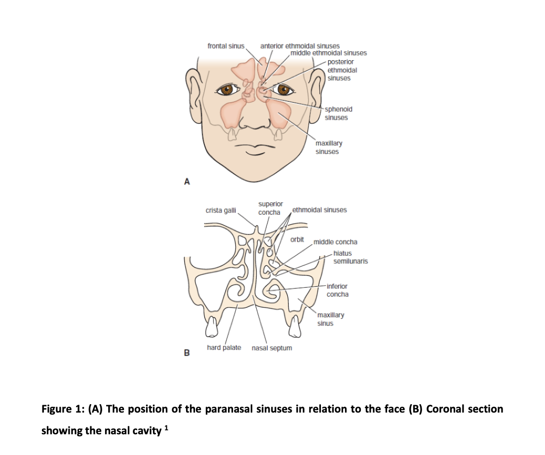
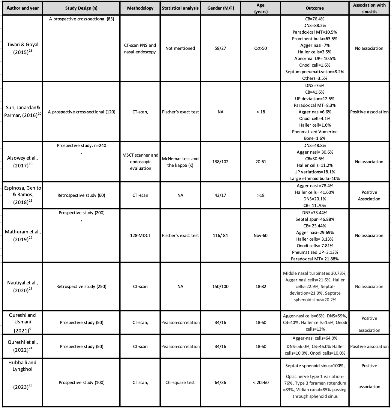
The nasal and paranasal sinus (PNS) structures (Figure 1) are important regions of distinct interest in various medical fields including the maxillofacial-surgery, and otorhinolaryngology. The PNS, which are carefully concealed, have repeatedly confused the anatomists and medical experts of the past years. Probably, because these structures are closely connected to many important organs of the body, for example, the brain, eyes, nose, and mouth, many unusual schemes about their purpose have been established for years. The sinuses were considered a mysterious area of a skull by previous anatomists. Historically, it was first recognized by ancient Egyptians then later contribution was made by Greek physicians. During Middle Ages, the anatomists Renaissance period-Leonardo-da-Vinci and Vesalius participated to make further enhancements to an understanding of complex anatomy1. The pathological changes within nasal and PNS are often of greater interest to radiologists than anatomical variations. But the anatomical variable features of the upper respiratory tract, that is nasal and PNS, are greatly significant because of their vital responsibility in the drainage pathway of the osteo-meatal-complex (OMC) and ventilatory sinus functioning. Thus, anatomical variants of the sino-nasal region elevate the risk of sinus-mucosal diseases. Furthermore, anatomical variations can affect the consequences and safety of surgical procedures performed in the sino-nasal region such as functional-endoscopic-sinus-surgery (FESS) 1-5. This review includes studies from 2013-2023 which were extracted from different databases. The aim of the review was on hidden anatomy of the nasal and paranasal sinus region, detected by computed tomography and highlighting the operative significance to enhance the surgical outcomes globally.
The diseases of the nasal cavity and PNS are among the most common pathologies which bump into the clinics of the ear, nose, and throat (ENT). Anatomic variations of the sinonasal region are also frequently observed, and they play a vital role in the dysfunctional drainage of sinus cavities. Computed tomography (CT imaging) is important to evaluate various pathologies of the nasal cavity and PNS. It allows us to assess the fine bony details related to the nasal cavity and PNS, anatomical variants, and disease progress of the sinonasal region. The CT imaging technique is now established as the overall best method for the evaluation of patients who are suspected of having complex anatomy and any aggressive lesion of the sinonasal region.
Sinonasal Framework
For a strong understanding of structural anatomy and variations of nasal and PNS regions, it is valuable to appreciate the various anatomical characteristics and relationships of these structures to their surroundings. The lateral wall of the nasal cavity contains several structural characteristics and recesses that are critical for the extensive understanding of PNS morphology, such as nasal turbinates which are three to four bony shelves, and the three nasal meatuses which are spaces situated below each nasal turbinate. The sphenoid and posterior ethmoid sinuses drain into the superior meatus. The middle nasal meatus present between the superior and inferior recesses receives sinus drainage from the frontal, maxillary, and anterior ethmoid.
The lower inferior nasal meatus receives drainage from the nasolacrimal duct. The sickle-shaped thin bony structure called uncinate process is a part of the ethmoid bone, which is sheathed by mucoperiosteum, situated medial and lateral to the ethmoid infundibulum and middle turbinate respectively. There is a pyramidal space called the ethmoid infundibulum, which helps in facilitating the drainage of the frontal, anterior ethmoid, and maxillary sinuses. The uncinate process (UP) and ethmoid bulla have a gap in between called semilunar hiatus, that empties the ethmoid infundibulum. The osteo-meatal complex (OMC) is situated near the middle turbinate laterally possess openings of various sinuses like maxillary, ethmoid, and frontal. This is a collection of various anatomical features located within the middle nasal meatus like the uncinate process, middle meatus itself, infundibulum, frontal, maxillary, ethmoid sinuses, and anterior ethmoid air cells along with sinus ostia6-9.
Sinonasal Anatomical Variants and Sinusitis
Anatomical variations of sinonasal area were very common, and many authors classified these variants into four familiar groups: nasal septum variations for instance deviated nasal septum (DNS), middle turbinate variations like concha bullosa (CB), uncinate process (UP) and ethmoidal anatomical variations10-12. The DNS was the most common anatomical variant found in patients (48.8%) with acute/chronic or recurrent sinusitis. The structures like agger-nasi-cells and CB were almost similarly frequent (30.7%), and infraorbital cells or Haller cells were noticed in 11.5%. The UP anatomical variants were noticed in 18.2%, and the large prominent ethmoid bulla was seen in 10%.10 The significance of anatomic structural variations requires attention in that way that these variations can impair the normal sinus drainage process of the associated sinus, which could result in various pathologies like inflammatory sinus diseases such as sinusitis. Generally, the anatomy varies from individual to individual globally and anatomical structural variations are harmless and are not diseases however, it may exist as an incidental finding in people with sinus infections10-12. The mucosa lining of the PNS is prone to infection and inflammation. Previously, Hippocrates specified that “In a person having a painful spot in the head, with intense headaches, pus or fluid running from the nose removes the disease” which may be described as sinusitis. It is the inflammation/swelling of the mucosal coating of paranasal sinuses (PNS), and the gaps that secrete mucus which is needed for the whole nasal passage to work effectively. This inflammatory condition called sinusitis is one of the most observed illnesses of the upper respiratory tract. The prevalence of sinusitis noticed globally says that it affects one in seven adults resulting in approximately fifty million individuals being diagnosed as patients of sinusitis annually13-15. This disease is increasing dramatically in epidemic proportions all over the globe. The conditions like chronic sinusitis and recurrent sinusitis have been recognized to influence adversely health-related quality of life within the community16.
Computed Tomography Significance for Anatomical Variants Evaluation
With the invention of FESS and CT, significant attention has been directed toward the anatomy of the PNS region. Detailed knowledge of anatomical variations in the PNS area is of critical value for surgeons executing endoscopic sinus surgery as well as for radiologists that are involved in the preoperative work-up of the patients17-19. Harmless anatomical variations are considered normal and can be present in any individual. The importance of anatomical variants can’t be ignored as the presence of various anatomical variants produces a diversity of relationships to the structure in which they lie. The Sino nasal cavities possess a multitude collection of anatomic variants, few of which are significantly common and observed in most people. In 2015 it was revealed that there was no noteworthy difference in the prevalence of variable anatomical features of PNS or nasal cavity between patients with mild sinus disease progress versus moderate to severe sinus mucosal disease. However, evaluation of diverse anatomical variants in every routine CT-PNS obtained for rhinosinusitis is of significant worth except surgery is aimed. Importantly, for cases who are intending to undergo FESS or other skull base surgery, however, the caregivers need to be aware of variants, such as onodi cells, supraorbital cells, infraorbital Haller cells, and many others. There are chances of a higher rate of surgical complications if variants are overlooked19-26.
In the last few years, the most useful sinus surgical technique called functional endoscopic sinus surgery (FESS) has developed a gold standard, especially in the treatment of recurrent or chronic rhinosinusitis. Therapeutic outcomes rely on the preoperative evaluation of the patients. The radiologic CT scanning of the PNS displays appropriate sensitivity and good specificity for the early diagnosis of pathologies like rhinosinusitis and the detection of anatomical variations of the sinonasal region. And the pre-FESS evaluation workup is globally recommended to provide detailed visualization of normal, variable, and pathological structures of the sinonasal region. Surprisingly, one study reported that variations are common in chronic rhinosinusitis, and which are possibly associated with localized chronic rhinosinusitis as compared to diffuse one which involves a majority of the sinuses. Further stated that the anatomical variations are not associated with the incidence of polyps. Moreover, computed tomography helps to reflect the disease severity, also, to some extent, the symptoms. Combination of medical history, physical examination, and in addition to it CT may increase the accuracy of diagnosing various sinus pathologies for instance rhinosinusitis27-29.
With the invention of new surgical modalities like FESS and new radiologic investigations like CT scanning, significant consideration has been directed toward the morphology of the nasal and PNS area. Comprehensive knowledge of anatomical variations in the PNS area is of crucial value for caregivers executing sinus surgery along with radiologists that are concerned with the patient’s preoperative work-up. The diseases of the nasal cavity and PNS are among the most common pathologies which bump into the clinics of the ear, nose, and throat (ENT). Anatomic variations of the sinonasal region are also frequently observed, and they play a vital role in the dysfunctional drainage of sinus cavities, generally resulting in acute sinusitis followed by chronic sinusitis30-36. A study concluded that variants were highly prevalent on computed tomography of nasal and paranasal structures and the frequently observed variant was a septal deviation most of the patients had more than one variant present, also those subjects were more at risk to develop sinusitis37. Surprisingly similar studies were observed by another author who observed septal deviation as the most common anatomical variant and the occurrence of multiple anatomical variations in most of the patients. Moreover, in contrast to other studies, the author reported that the prevalence of anatomical variations does not describe the establishment of disease progress but may predispose it to operative complications. Hence, radiologists play a crucial role to identify and detect anatomical variants to provide a clear pathway to surgeons and reduce the chances of operative complications38. Interestingly, one study stated that nasal and paranasal symptoms in SARS-CoV-2 positive subjects are usually rare when compared to the lower respiratory symptoms. But supporting the practice of computed tomography for the evaluation of nasal and paranasal structures for proper visualization39. The paranasal structures are a group of pneumatized spaces developed as an extension of the nasal cavities, eroding the adjacent bony structures. According to the research, some regions present a high risk for trauma and significant intraoperative complications, with the frontal and ethmoid sinuses being most frequently affected. Anatomical variations, in correlation with their inherent conditions, are added to surgical risks so the knowledge of these vital structures is critical for endoscopic surgeons as well as for radiologists contributing to the preoperative evaluation of the patients, to avoid therapeutic letdown and complications. The gaining of an excellent definition of the sinonasal anatomy for a preoperative evaluation can be done by computed tomography which is the gold standard in the study of complicated structures, for providing accurate information on soft tissues, bony regions, and the presence of air cells, thus characterizing a highly sensitive method of imaging39-43.
Highlights of Nasal and Paranasal Anatomical Variations From 2015-2023
Nowadays increasing interest in FESS has put the characteristic distinguishable nasal and paranasal anatomy in the spotlight. Now in recent years, research about the correlation between anatomic variations of sinonasal region and sinusitis has been increasing in order to determine the clinical significance of anatomic variants to prevent sinus mucosal pathologies, but still, it is a matter of discussion. It is argued by several authors that some anatomic variants of sinonasal territory can cause sinus drainage obstruction, therefore becoming the reason for various sinus mucosal infections. Presently the modality of choice for the evaluation of nasal structures, sinuses along with its nearby relations, is computed tomographic imaging. This imaging technique displays accurate information on both bony and soft tissue details along with the extent of disease within paranasal sinuses including adjacent structures. In contrast to ordinary radiography, CT imaging can undoubtedly display the fine bony anatomy and variations of the sinuses and OMC channels. The research showed that the significance of anatomical variants of the sinonasal region is still controversial. The majority of authors believe that various anatomic variants can cause some way for the occurrence of recurrent rhinosinusitis.
Therefore, it can be summarized that the use of computed tomographic scanning is the best approach to visualize clinically significant anatomy and additionally the disease diagnosis of sinuses and nasal areas. Surgeons learn from their mentors the important instruction of sinus surgeries to spare as much as possible and remove as much as required. The choice of investigation for the patients who are expected for surgical interventions like FESS is computed tomography. It will assist the surgeons to visualize the anatomic structural features residing within the nasal and paranasal areas. Therefore, it is labeled as a ‘‘ROAD MAP OF FESS”. The paranasal sinuses are critically important having complex anatomical variations, and variable anatomical relations, hence CT scan is advisable for all patients who are undergoing surgical intervention, to prevent dreadful complications39-40. The detailed workup of some previous studies from 2015 to 2023 is compiled in Table-1.
The prevalence of anatomical variations is quite common globally and septal deviation is the most frequent variant and multiple variations are more commonly observed, importantly which makes them more vulnerable to sinus pathologies. Researchers across the globe have appreciated the use of computed tomography of a sinonasal region in every patient of sinonasal pathology to provide a clear road map to healthcare professionals for better therapeutic strategies, surgical outcomes, and enhancing the quality of life within the community.
No conflicts of interest to declare.
All authors equally worked on the literature search and drafting of the manuscript.
- Snell RS. Snell’s Clinical Anatomy. Wolters kluwer India. 2018,9th Ed, pp: 643.
- Schuez I, Alt KW. Leonardo da Vinci and dental anatomy. Journal of Anatomy. 2022 ;240(2):183-196. doi: https://doi.org/10.1111/joa.13561
- Vaid S, Vaid N. Sinonasal Anatomy. Neuroimaging Clinics. 2022;32(4):713-734. doi: https://doi.org/10.1016/j.nic.2022.07.007
- Qureshi MF, Usmani A, Mehwish A, Mahar Y, Khattak MA, Ahmed RR. Anatomical Variations of Rhinogenic Headache and Its Relation with Sinusitis: A Computerized Tomography (CT) Scan Study.Pak J Med Dent. 2022;11(3): 37-43. doi: 10.36283/PJMD11-3/007
- Gandhi KR, Patil ST, Kumar B, Patel M, Chaware P. Study of frontal and ethmoid sinus of sinonasal complex along with olfactory fossa: anatomical considerations for endoscopic sinus surgery. Anat Cell Biol. 2023,1-6. doi: 5115/acb.22.230
- Cappello ZJ, Minutello K, Dublin AB. Anatomy, Head and Neck, Nose Paranasal Sinuses. In: StatPearls. StatPearls Publishing, Treasure Island (FL); 2022.
- Abdalla MA. Human maxillary sinus development, pneumatization, anatomy, blood supply, innervation and functional theories: An update review. Siriraj Medical Journal. 2022;74(7):472-479. doi: https://doi.org/10.33192/Smj.2022.56
- Qureshi MF, Usmani A. Clinically significant variation of paranasal sinuses on CT-scan. JBUMDC. 2020;10(2):152-157. Doi: https://doi.org/10.51985/JBUMDC2019138
- Qureshi MF, Usmani A. A CT-Scan review of anatomical variants of sinonasal region and its correlation with symptoms of sinusitis (nasal obstruction, facial pain and rhinorrhea). Pak J Med Sci. 2021;37(1):195-200. doi: https://doi.org/10.12669/pjms.37.1.3260
- Alsowey AM, Abdulmonaem G, Elsammak A, Fouad Y. Diagnostic performance of multidetector computed tomography (MDCT) in diagnosis of sinus variations. Pol J Radiol, 2017; 82: 713-725. doi: https://doi.org/10.12659/PJR.903684
- Papadopoulou AM, Chrysikos D, Samolis A, Tsakotos G, Troupis T. Anatomical variations of the nasal cavities and paranasal sinuses: a systematic review. Cureus. 2021;13(1). doi: 10.7759/cureus.12727
- Devaraja K, Doreswamy SM, Pujary K, Ramaswamy B, Pillai S. Anatomical variations of the nose and paranasal sinuses: A computed tomographic study. Indian J Otolaryngol Head Neck Surg. 2019;71: 2231-2240.doi: https://doi.org/10.1007/s12070-019-01716-9
- Rajashree AF, Deepthi P, Viswanatha B. Impact of concha bullosa on osteomeatal complex drainage and septal deviation. Res Otolaryngol. 2018;7(1):1-4.
- El-Din WA, Madani GA, Fattah IO, Mahmoud E, Essawy AS. Prevalence of the anatomical variations of concha bullosa and its relation with sinusitis among Saudi population: a computed tomography scan study. Anat Cell Biol 2021;54(2):193-201. Doi: https://doi.org/10.5115/acb.20.247
- Zahedi FD, Yaacob NM, Wang DY, Abdullah B. Radiological anatomical variations of the lateral nasal wall and anterior skull base amongst different populations: A systematic review and meta‐analysis. Clinical Otolaryngology.2023;48:271–285.Doi: https://doi.org/10.1111/coa.13975
- Orlandi RR, Kingdom TT, Smith TL, Bleier B, DeConde A, Luong AU, Poetker DM, Soler Z, Welch KC, Wise SK, Adappa N. International consensus statement on allergy and rhinology: rhinosinusitis 2021. International forum of allergy & rhinology 2021;11(3), 213-739. Doi: https://doi.org/10.1002/alr.22741
- Kato A, Peters AT, Stevens WW, Schleimer RP, Tan BK, Kern RC. Endotypes of chronic rhinosinusitis: relationships to disease phenotypes, pathogenesis, clinical findings, and treatment approaches. Allergy. 2022;77(3):812-26. doi: https://doi.org/10.1111/all.15074
- Sedaghat AR, Kuan EC, Scadding GK. Epidemiology of chronic rhinosinusitis: prevalence and risk factors. The Journal of Allergy and Clinical Immunology: In Practice. 2022;10(6): 1395-403.doi: https://doi.org/10.1016/j.jaip.2022.01.016
- Tiwari R, Goyal R. Study of anatomical variations on CT in chronic sinusitis. Indian J Otolaryngol Head Neck Surg. 2015; 67:18-20.
- Suri N, Janardan T, Parmar H. Correlation of anatomical variations of Paranasal sinuses and Chronic Rhinosinusitis. Int Arch Integrated Med. 2016;3(12):84-8.
- Espinosa W, Genito R, Ramos RZ. Anatomic variations of the nasal cavity and paranasal sinus and their correlation with chronic rhinosinusitis using Harvard staging system. J Otolaryngol ENT Res. 2018;10(4):190-193.
- Mathuram AC, Aiyappan SK, Agarwal S, Raveendran NH, Valsala VS. Assessment of Sinonasal Anatomical Variants using 128-Slice MDCT in Patients with Chronic Rhinosinusitis. Radiology. 2019;4(2):120-126.
- Nautiyal A, Narayanan A, Mitra D, Honnegowda TM. Computed tomographic study of remarkable anatomic variations in paranasal sinus region and their clinical importance-A retrospective study. Annals of Maxillofacial Surgery. 2020;10(2):422-428. doi: 10.4103/ams.ams_192_19
- Qureshi MF. Anatomical Variations of Rhinogenic Headache and Its Relation with Sinusitis: A Computerized Tomography (CT) Scan Study. PJMD. 2022;11(3):37-43. Doi: https://doi.org/10.36283/pjmd.v11i3.1694
- Hubballi RK, Lyngkhoi BL. Analysis Of The Anatomical Variations Of The Sphenoid Sinus In Patients With Chronic Rhinosinusitis. Indian J Otolaryngol Head Neck Surg. 2023:1-8. Doi: https://doi.org/10.1007/s12070-022-03385-7
- Khalid AB, Siddiqa A, Haider SI, Alam G, Butt NA. Anatomical Variations on Routine CT Scans Observed in the Paranasal Sinuse. PJMHS. 2022;16(03):731-733. Doi: https://doi.org/10.53350/pjmhs22163731
- Debnath J, Maurya V, Sharma V. Pre-FESS Imaging of Paranasal Sinuses and Nasal Cavity: Using Multi-detector Computed Tomography (MDCT) in Understanding Normal Anatomy and Anatomical Variations: Tips and Tricks. Indian J Otolaryngol Head Neck Surg. 2022:1-9.
- Liu L, Chen Q, Pan M, Yang Y. Roles of Anatomical Abnormalities in Localized and Diffuse Chronic Rhinosinusitis. Indian J Otolaryngol Head Neck Surg. 2023; 10:1-7.
- Reddy UD, Dev B. Pictorial essay: Anatomical variations of paranasal sinuses on multidetector computed tomography-How does it help FESS surgeons?. Indian Journal of Radiology and Imaging. 2012 Oct;22(04):317-324.
- Mokhasanavisu VJ, Singh R, Balakrishnan R, Kadavigere R. Ethnic variation of sinonasal anatomy on CT scan and volumetric analysis. Indian J Otolaryngol Head Neck Surg. 2019; 71:2157-2164.
- Dasar U, Gokce E. Evaluation of variations in sinonasal region with computed tomography. World journal of radiology. 2016;8(1):98-108. doi: 4329/wjr.v8.i1.98
- Marino MJ, Riley CA, Wu EL, Weinstein JE, Emerson N, McCoul ED. Variability of paranasal sinus pneumatization in the absence of sinus disease. Ochsner Journal. 2020;20(2):170-175.
- Shokri A, Faradmal MJ, Hekmat B. Correlations between anatomical variations of the nasal cavity and ethmoidal sinuses on cone-beam computed tomography scans. Imaging Science in Dentistry. 2019;49(2):103-113.
- Nikkerdar N, Eivazi N, Lotfi M, Golshah A. Agreement between cone-beam computed tomography and functional endoscopic sinus surgery for detection of pathologies and anatomical variations of the paranasal sinuses in chronic rhinosinusitis patients: a prospective study. Imaging science in dentistry. 2020;50(4):299-307. doi: 5624/isd.2020.50.4.299
- Hadi HH, Al-Bayati HA, Al-Gazali SS. Prevalence of normal anatomical variations in the region of paranasal sinuses in patients with chronic rhinosinusitis. Med J Babylon. 2018;15(3):243-250. Doi: 10.4103/MJBL.MJBL_71_18
- Vora S, Patel M, Shah B, Patel R. Prospective cross sectional study on anatomical variation with special emphasis on critical anatomical landmark in patients undergoing multi detector computed tomography of paranasal sinuses: Anatomical variation in CT PNS. GAIMS Journal of Medical Sciences. 2023; 3:13-18. Doi: https://gjms.gaims.ac.in/index.php/gjms/article/view/51
- Al-Ani RM, Khalaf GM. Prevalence of sinonasal anatomical variations and their effect on chronic rhinosinusitis in Al-Ramadi Teaching Hospital, Ramadi City, Iraq. Muthanna Medical Journal. 2021;8(1):35-43.
- Devareddy MM, Devakar S. Evaluation of anatomical variations in nose and paranasal sinuses by using multidetector computed tomography. Int J Contemp Med Surg Radiol. 2019;4(3):C146-C151. DOI: http://dx.doi.org/10.21276/ijcmsr.2019.4.3.32
- Kalyankar AG, Sukre SB, Kulkarni P. Anatomical study of variant paranasal sinuses in accordance with its clinical significance by CT scan: Pre-COVID-19 period. International Journal of Anatomy and Physiology. 2022;11(1):1-6.
- Shiekh Y, Wani AH, Khan AJ, Bhat MI. Anatomical Variations of Paranasal Sinuses-A MDCT Based Study. Int Arch Intern Med. 2019; 6:300-306.
- AB R, HM F, EF AK, AM S. Anatomic variations of para-nasal sinuses in patients undergoing CT scan: spectrum, prevalence and implications. Benha Journal of Applied Sciences. 2022;7(3):89-99.
- Dawood SN. Normal anatomic variants of paranasal sinus region studied by computed tomography. Zanco J Med Sci. 2020;24(2):187-196.
- Adeel M, Rajput MS, Akhter S, Ikram M, Arain A, Khattak YJ. Anatomical variations of nose and para-nasal sinuses; CT scan review. J Pak Med Assoc. 2013;63(3):317-319.
This is an open-access article distributed under the terms of the CreativeCommons Attribution License (CC BY) 4.0 https://creativecommons.org/licenses/by/4.0/